
Since the 1980s, minimally invasive approaches have played an important role in general surgery as an alternative to traditional large incision open surgery. It can also be used for many surgical techniques within the thoracic cavity, ranging from cardiovascular, oncological, pulmonological, even spinal operations. Three in four prostatectomies in the United States are performed robotically-assisted. Biomedical innovation and technological advancement have seen surgical techniques in minimally invasive approaches rapidly grow. But what is it, and how exactly does it work?
One of the most common, if not the most common robotically-assisted surgical system is called Da Vinci. It’s produced by Intuitive, a publicly traded company based out of Sunnyvale, California. Since its FDA approval in 2000, Da Vinci has significantly helped accuracy and patient well-being. There are three Da Vinci systems: Da Vinci X, the standard model; Da Vinci Xi, the most advanced model; and Da Vinci SP, the single-port variant. The use of the system is dependent on the type of surgery being performed.
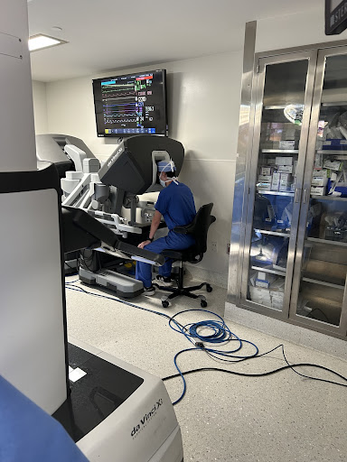
Let’s use the procedure I witnessed in the cover photograph. The patient was diagnosed with coronary artery disease, or plaque blockage in their coronary arteries (known as atherosclerosis). If the coronary arteries are blocked, the heart is starved of oxygen and multiple severe complications can occur. The solution is a surgical operation called a coronary artery bypass graft, or CABG. Depending on the condition of the heart, one, two, three, or even four grafts would have to be made to “bypass” the affected artery by connecting it externally to the artery and allowing blood to flow around the blocked area. A cardiac catheterization confirms the exact location of the blockage to allow the surgeon to create the bypass in the perfect location.
After my dad, a cardiac anesthesiologist, administered anesthesia, the surgeon created three incisions, or ports, for three of the four arms to access the heart. Two of these arms were used to maneuver around the area, while the third was a camera to both illuminate and allow viewing of the site. Once the arms were in place, the surgeon de-scrubbed and sat at the surgeon’s console. In this case, it was a single bypass graft, so only one artery was taken to bypass the blockage (the internal mammary artery, running through the left side of your chest). In cases requiring more than one bypass, the saphenous vein is taken from the left leg and is usually cut into multiple pieces to create more grafts.
Using a stereoscopic video, the same imagery that gives VR headsets a 3D illusion, the surgeon used the two arms to carefully remove the artery from the chest wall. A combination of two maneuverable handles and pedals allowed the surgeon to perform this difficult task with ease. Surges of controlled electricity ran through the metal tips of the arms, cauterizing the artery. This allocated not only clean removal from the chest wall but it also allowed the surgeon to burn off any bleeding that may occur from surrounding blood vessels. A built-in microphone allowed the surgeon to speak to the physician’s assistant who stood near the table through a speaker system. Once the internal mammary artery was removed, the surgeon then scrubbed back in to make a small incision, around two inches, in the left side of the chest and made the bypass. That was it!
The most significant difference of this operation as opposed to standard approaches to CABG is the ludicrous difference in incision sizes. The minimally invasive approach used three very small incisions around the rib cage, as well as one two-inch incision on the left side of the chest. However, the traditional open heart approach uses a very large eight to twelve inch incision, going all the way down the breastbone and completely separating it.
When someone has a tumor or lesion in their lungs, a biopsy is performed to remove the object. Another robotic system made by Intuitive, called Ion, creates a minimally invasive approach to peripheral lung bronchoscopies with a personalized passage system made by their data system.
It begins with Intuitive’s PlanPoint software. PlantPoint uses a CT scan to 3D generate the patient’s airway trees within the lungs. When the lesion is identified, PlanPoint automatically generates a path through the lung to allow access to the lesion. This is done by its deep segmentation capabilities powered by machine learning. Then, based on their distance to the lesion, structures of pleura or fissures are generated. This helps the operator visualize what they will be seeing when they perform the biopsy. If needed, PlanPoint can create alternative routes to the lesion.
Once the bronchoscopy is underway, the operator will use the control setup, which looks like a small white laptop, to guide the ultrathin robotic catheter through the pre-planned path toward the lesion. The catheter has a small 3.5 mm outer diameter and a 2.0 mm working channel. It can maneuver 180 degrees in any direction around tight turns, allowing access to all 18 segments of the lung. It is equipped with a fiberoptic sensor which measures the full shape of the catheter, allowing real-time exact location and geometric information.
When the lesion is reached, the catheter is locked in place. Ion’s Flexision Needle, designed to pass through the catheter, deploys into the target location on a straight path. The biopsy market feature allows the tracking of multiple biopsy attempts, helping the operator visualize different biopsy needle trajectories. If confirmation of a tissue sample is available, the operator can redirect upcoming biopsy attempts to the target area. This is referred to as a cloud biopsy.
Patients who undergo minimally invasive techniques opposed to traditional open methods have much quicker recovery times. They also have a smaller chance of postoperative complications due to a smaller operational area. Minimally invasive approaches have significantly helped both surgeons and patients alike to bring them to full health, and new developments in these systems pave the way for medicine to be as advanced as possible.